Introduction

In assisted reproductive technologies (ART), time-lapse incubation has proven to be a revolutionary approach to observing embryo development. In this method, embryologists can monitor the growth of the embryo through continual images in a closed environment, revealing critical developmental markers without disrupting the embryo’s environment.

What is Time-Lapse Incubation?
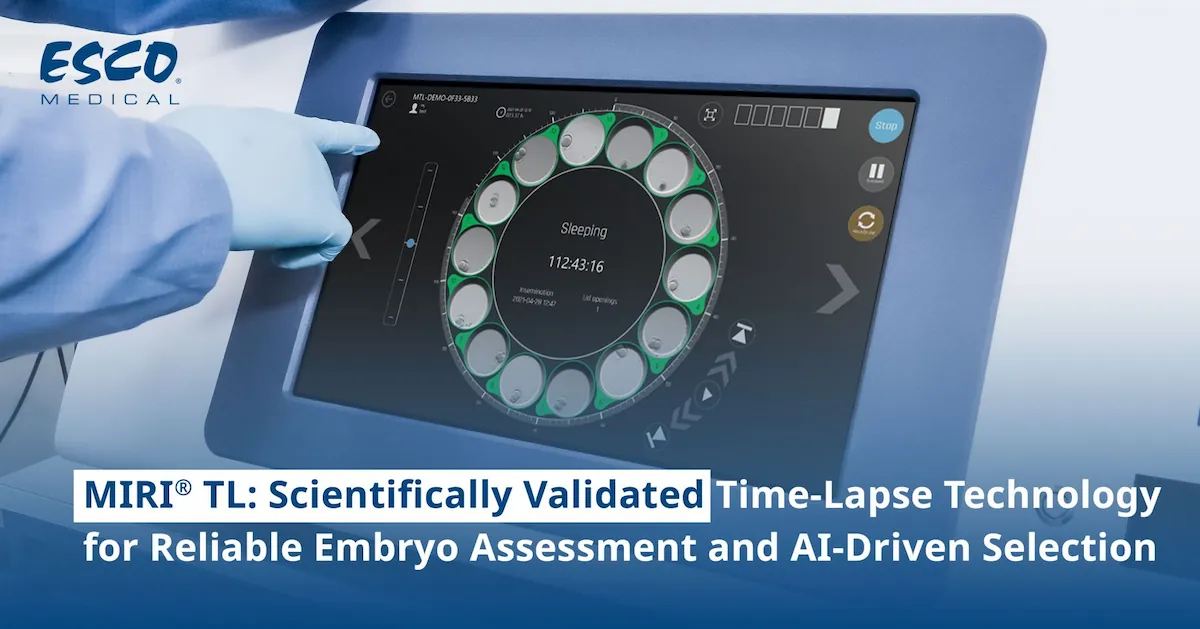
Time-lapse technology, done with an incubator like Esco’s MIRI® TL Incubator, enables non-invasive observation of embryos throughout their development. It involves placing embryos in a stable culture environment where cameras capture images at consistent intervals. These images are then assembled to make a time-lapse video by which embryologists can observe each phase carefully. Through this continuous monitoring they get to learn some of the important developmental steps—information that aids in selecting the most viable embryos for transfer in IVF procedures.
Why is Time-Lapse Incubation Important?
Another benefit of time-lapse incubation is that it provides information on the development steps live and in real-time. In comparison to conventional methods which involve the frequent evaluation and often transfer of embryos back to the incubator, this method records the entire culture period within the incubation box. This real-time tracking reduces the frequency of manual movement of the embryos, thus preserving the environment that is crucial in the growth of the embryos.
Furthermore, as ART continues to advance, there is increasing demand for precision and non-invasive methods that ensure embryo health and viability. Time-lapse systems meet these demands by providing detailed insights that improve embryo selection accuracy, ultimately supporting better IVF outcomes and enhancing the quality of fertility care.


Problems in Time-Lapse Incubation

Challenges in Image Quality
One of the challenges here is getting high-quality images that enable one to identify the development milestones. Traditional imaging methods sometimes fall short, producing images that lack the resolution needed for detailed embryo assessment. Low-quality images reduce the visibility of certain features that help embryologists make proper evaluations for the viability of the embryo. This limitation can affect embryo selection since lower-quality images may hide important developmental details necessary to abstract embryo viability and health.
Issues with Undisturbed Incubation
An ideal incubation system would maximize time-lapse efficiency without requiring frequent human intervention. Frequent manual handling of embryos can be an unfavorable process because the manipulations lead to changes in temperature and other conditions that are required for the further development of embryos. Current systems aim to minimize these disruptions by automating image capture and ensuring stability, yet further improvements are needed to maintain an undisturbed environment continuously.

The Solution: Introducing MIRI® TL

MIRI® TL

What is MIRI® TL?

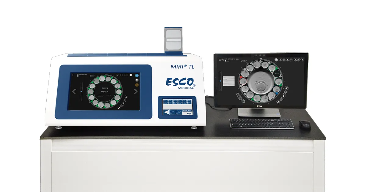
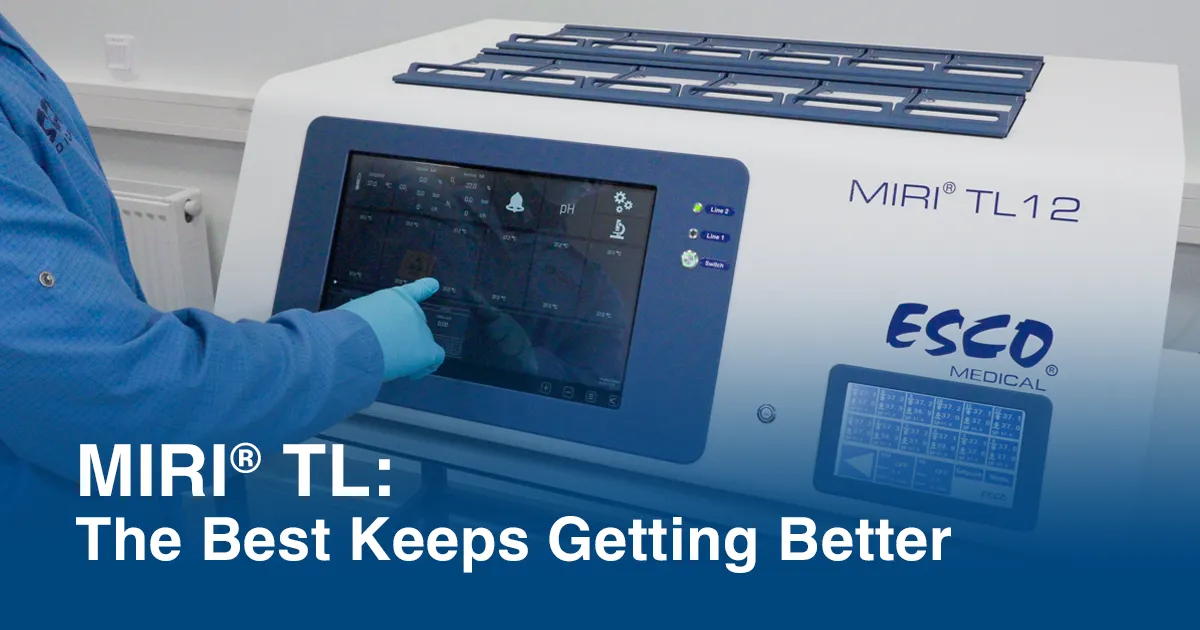
The MIRI® TL is a time-lapse incubator designed to solve the challenges embryologists face in monitoring embryo development. MIRI® TL has been developed with state-of-the-art imaging features; this enables embryologists to capture high-resolution images of embryos in a stable, controlled environment, allowing embryologists to observe developmental markers clearly and accurately.
MIRI® TL features best in the market temperature and gas system, showcasing ± 0.1°C temperature and ± 0.2% gas stability within the setpoint. Separate incubation chambers limit the number of times that embryos are moved, which, in turn, decreases contamination and increases environmental stability. This incubator combines innovative technology with reliability and is a helpful instrument to accomplish the highest possible success in ART.


Our MIRI® time-lapse incubator is now equipped with a new set of features to further revolutionize embryology.

New Features and Benefits

Enhanced Image Quality

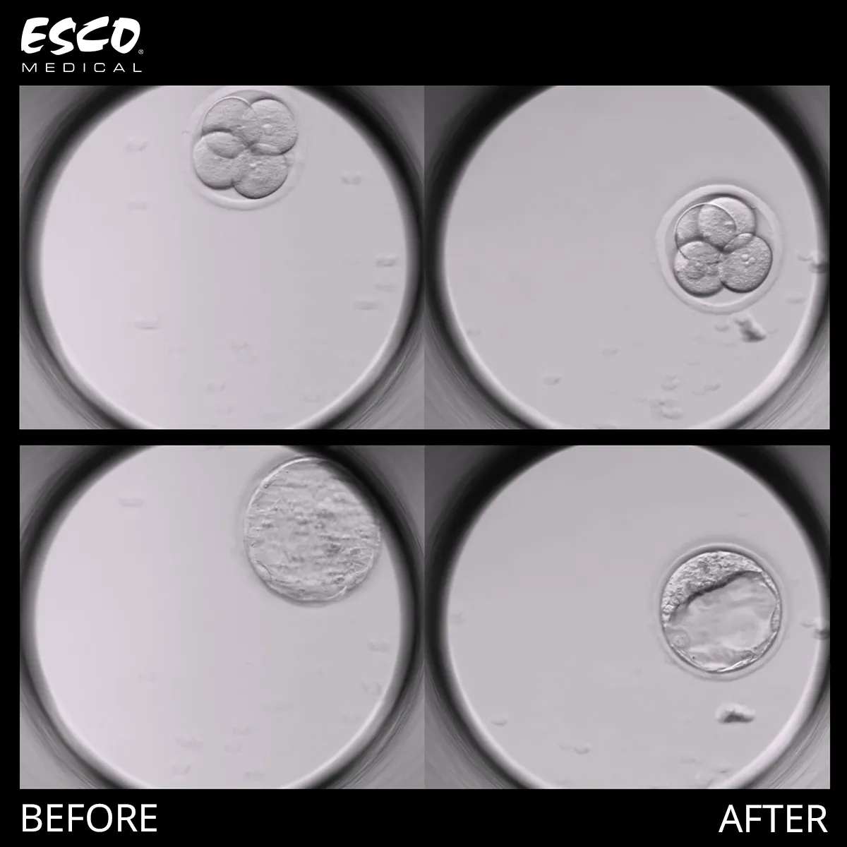
Monitor your embryonic development with remarkable clarity, thanks to the improved MIRI® time-lapse incubator. Superior image resolution and contrast enable embryologists to monitor cytoplasm features in better detail and detect abnormalities, such as:
Large perivitelline space
Dark zona pellucida
Presence of vacuoles and refractile bodies
MIRI® TL Before After

Esco Medical MIRI® time-lapse incubator image quality has always met the high standards required for precise embryological assessment, and now, with implemented enhancement, it is even better. This improvement further supports the distinction between normal and abnormal fertilization by enabling detailed visualization of pronuclei size and number. By Day 2 and Day 3, the system delivers exceptionally clear views of cell boundaries between blastomeres, facilitating accurate cell counts, evaluation of blastomere evenness, and identification of key morphological features such as vacuoles and multinucleation. The refined resolution enhances the ability to assess blastomere compaction at the morula stage and provides superior detail when evaluating the trophectoderm quality and inner cell mass thickness at the blastocyst stage, empowering embryologists with unparalleled clarity at every developmental milestone.

Undisturbed Embryo Development

Providing a highly stable environment with minimum need for manual intervention, the MIRI® TL time-lapse incubator will support embryologists in creating optimal growth conditions throughout the entire incubation period. Key factors include:
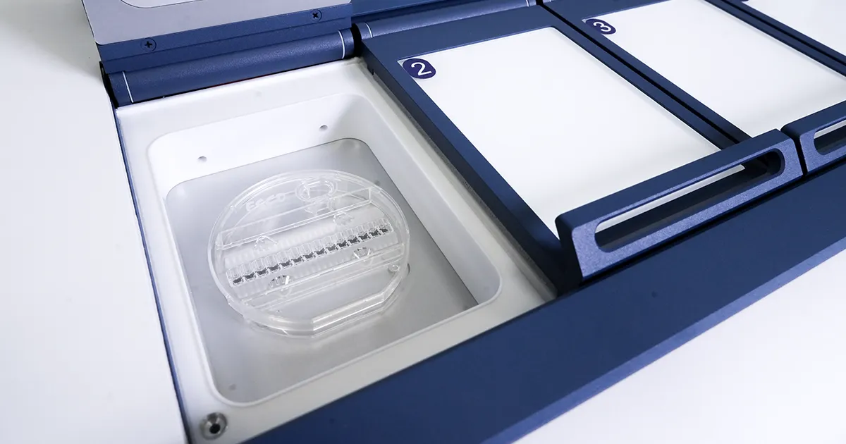
Individual chambers for each patient: The incubator prides itself on maintaining constant temperature and gas levels without needing to open all incubator’s chambers, which is crucial for early cleavage embryos that are sensitive to changes in the culture environment.
Unmatched gas and temperature system stability: Our proven, industry-leading gas system ensures unmatched stability and precision, maintaining optimal concentration and temperature levels with rapid recovery rates. This guarantees the most reliable environment for embryo development, giving embryologists complete confidence in their culture conditions.
Long-term stability: Embryos can be cultured in the same medium for five to six days, which will reduce exposure to potential disruptions and provide the suitable environment necessary for healthy development.
MIRI® TL CultureCoin®

This undisturbed environment is especially important for early-stage embryos, which are more prone to fluctuations in their surroundings than blastocysts.

The undisturbed culture environment supports ideal growth and the superior image quality aids monitoring with increased precision.

For inquiries visit https://www.esco-medical.com/contact-us.

References

https://www.esco-medical.com/products/time-lapse-embryo-incubator